Medical Imaging
Functional MRI (fMRI) Explained: Brain Mapping Before Neurosurgery
Reviewed by admin · Last updated June 13, 2026
Functional MRI, usually shortened to fMRI, is a specialized form of brain imaging that shows which regions of the brain are active while a patient performs specific tasks — rather than simply showing brain structure the way a standard MRI does. In Turkey’s leading neurology and neurosurgery centers, fMRI has become an important planning tool used before certain brain surgeries to help surgeons avoid damaging areas responsible for critical functions like speech, movement, or memory. This guide explains how the technology works and what patients can expect from the scanning process.
Rexalife is a medical consultancy. We help international patients access accredited imaging centers and neurosurgical teams in Turkey that offer fMRI as part of surgical planning — we do not perform imaging or interpret scans ourselves.
How Is fMRI Different From a Standard MRI?
A conventional MRI produces detailed, static images of brain anatomy — the shape, size, and structure of different regions, useful for identifying tumors, structural abnormalities, or areas of damage. Functional MRI, by contrast, detects changes in blood flow and oxygen levels that occur when a specific brain region becomes more active. Because active neurons demand more oxygenated blood, fMRI can effectively create a real-time map of which parts of the brain “light up” while a patient performs a particular mental or physical task during the scan — hence the description of fMRI as showing brain function rather than just structure, a distinction also relevant when comparing it to our overview of standard brain MRI.
Why Is fMRI Used Before Brain Surgery?
When a neurosurgeon needs to remove a brain tumor or treat epilepsy surgically, one of the biggest risks is inadvertently damaging brain tissue responsible for essential functions such as language, movement, or vision — tissue that can sit dangerously close to, or even within, the area requiring surgery. fMRI allows the surgical team to map exactly where these critical functional areas are located in that specific patient’s brain before the operation begins, since brain anatomy for functional regions varies somewhat from person to person, especially in patients who have had prior brain injury or whose tumor may have caused the brain to reorganize function around it.
What Tasks Do Patients Perform During the Scan?
During an fMRI session for surgical planning, patients typically perform specific tasks while lying in the scanner — this might include finger tapping to map the motor cortex, naming objects shown on a screen to map language areas, or simply reading and listening to identify areas involved in comprehension. The scan captures brain activity throughout these tasks, and specialized software then overlays this functional activity map onto the structural MRI images taken during the same session, giving surgeons a combined picture of both anatomy and function to guide their surgical approach.
What the Scanning Session Actually Involves
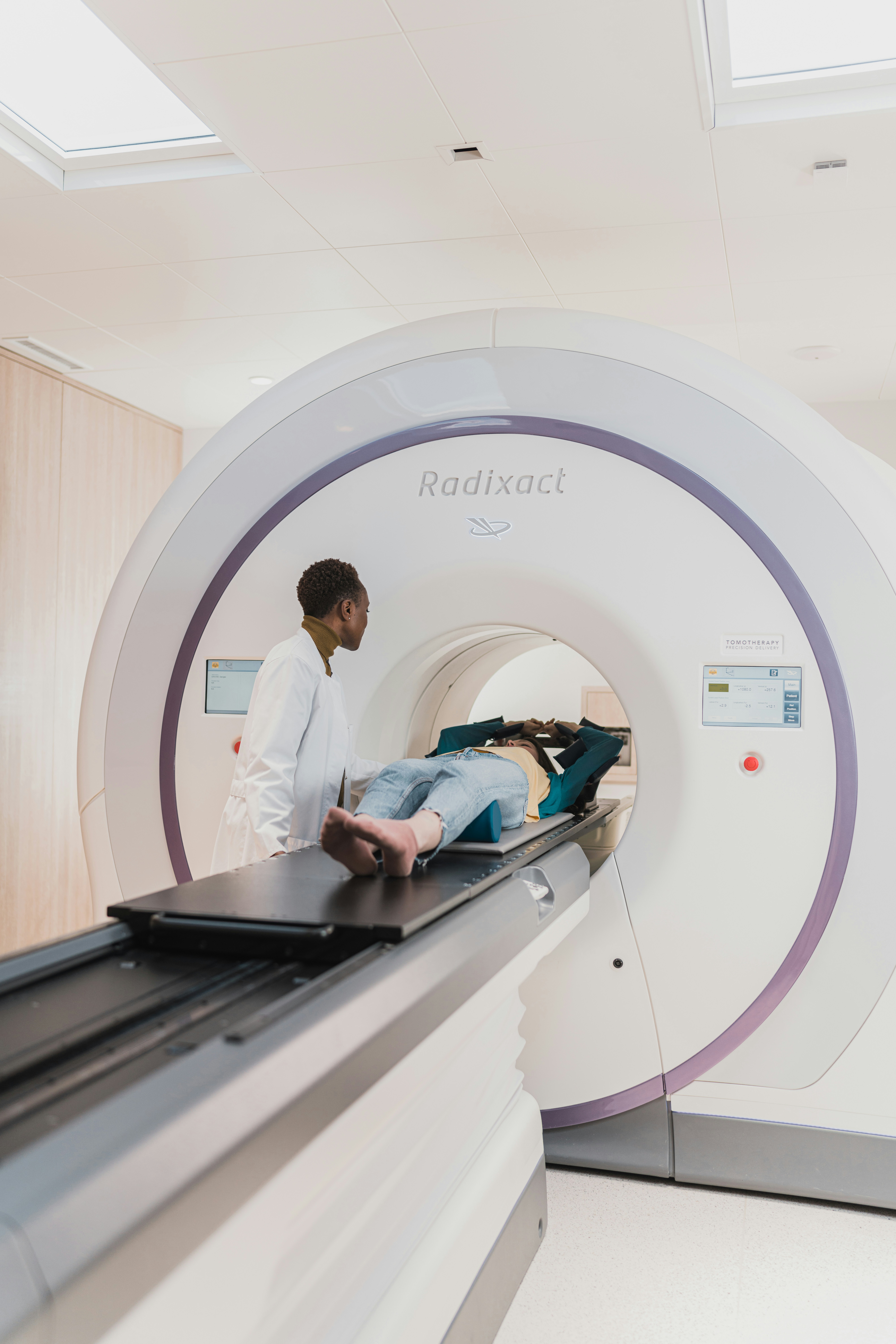
An fMRI session for surgical planning generally takes longer than a standard MRI, often 45 minutes to over an hour, because it includes both structural imaging and multiple functional task sequences. Patients lie still inside the MRI scanner, much like our overview of how long an MRI scan takes describes for conventional scans, but are asked to actively perform tasks at specific points rather than simply remain still throughout. Communication with the scanning team via an intercom and sometimes a small mirror or screen inside the scanner allows patients to see instructions and respond appropriately during the functional sequences.
Who Typically Needs fMRI?
fMRI for surgical planning is generally reserved for patients undergoing surgery for brain tumors located near functionally important regions, patients being evaluated for epilepsy surgery where identifying the seizure focus relative to critical brain areas is essential, and occasionally patients undergoing certain other neurosurgical procedures where precise functional mapping materially changes the surgical approach. It is not a routine test for general health screening — it is a targeted, purpose-specific tool used as part of a defined surgical workup, distinct from screening-oriented imaging discussed in our guide to full body MRI.
Combining fMRI With Other Pre-Surgical Imaging
fMRI is often used alongside other advanced imaging techniques as part of a complete pre-surgical picture, including diffusion tensor imaging to map white matter tracts — the brain’s structural “wiring” — which is covered in more detail in our guide to MRI spectroscopy and tractography. Combining these techniques gives the neurosurgical team both a functional map of what different brain regions do and a structural map of how they connect to each other, substantially reducing the risk of unexpected functional deficits after surgery.
Choosing the Right Center for fMRI in Turkey
Because fMRI for surgical planning requires specialized software, a technologist experienced in running functional task paradigms, and a radiologist or neurologist skilled in interpreting the combined functional and structural data, this is a service best sought at major neurosurgical centers in cities like Istanbul or Ankara rather than a general imaging clinic. Our broader overview of the best hospitals in Turkey for MRI, CT, and advanced diagnostics is a useful starting point when evaluating which center has genuine experience with this specific technique.
What Happens After the Scan
Once the fMRI data is collected, a specialized team — often including a neuroradiologist and sometimes a neuropsychologist — processes and reviews the functional maps alongside the structural images, producing a combined visualization that the neurosurgical team uses directly during surgical planning meetings. This analysis takes time, and results are not typically available immediately after the scan itself; patients should expect a review period of several days before the full report is ready and discussed with the referring neurosurgeon, who will explain what the findings mean specifically for the planned procedure.
Combining fMRI Findings With the Surgical Plan
The functional map produced by fMRI becomes one input among several that the neurosurgical team weighs when finalizing the surgical approach, alongside structural imaging, the patient’s neurological examination findings, and sometimes intraoperative monitoring techniques used during the surgery itself to further confirm functional boundaries in real time. Understanding that fMRI informs, rather than single-handedly dictates, the surgical plan helps patients appreciate why the pre-operative consultation with the neurosurgeon remains essential even after a detailed functional map has been produced.
Patient Anxiety and the Scanning Environment
Because fMRI sessions are longer than standard MRI and require active task performance, some patients feel more anxious about the process. Centers experienced in fMRI typically explain each task clearly beforehand, allow a practice run of simpler tasks before the actual recorded sequences begin, and maintain constant communication through the intercom throughout, which meaningfully reduces anxiety for most patients once they understand exactly what will be asked of them.
What to Bring to Your fMRI Appointment
- Referral letter and relevant prior imaging
- A list of any implants or metal devices in your body
- Comfortable clothing without metal fastenings
- A support person, if permitted, for reassurance
It’s worth noting that fMRI results are always interpreted within the broader context of your specific surgical case by the neurosurgical team, rather than viewed as a standalone diagnostic report. If you have questions about how your particular scan results will influence your surgical plan, your neurosurgeon is the right person to walk through this with you in detail before the operation date is confirmed.
How Rexalife Helps
We help international patients whose neurosurgeon has recommended fMRI as part of surgical planning connect with accredited centers in Turkey that have genuine, verified experience running these specialized scans, and coordinate the logistics between your imaging appointment and your surgical team. We do not perform imaging, interpret scans, or make surgical recommendations — that responsibility remains with your neurosurgical team throughout.
Frequently Asked Questions
What is the difference between fMRI and a regular brain MRI?
A regular MRI shows brain structure and anatomy, while fMRI shows which brain regions are active during specific tasks by detecting changes in blood flow, helping map brain function rather than just structure.
Why do surgeons use fMRI before brain surgery?
fMRI helps surgeons identify exactly where critical functions like speech, movement, or memory are located in an individual patient’s brain, reducing the risk of damaging these areas during surgery.
What do patients do during an fMRI scan?
Patients perform specific tasks inside the scanner, such as tapping fingers, naming objects, or reading, while the machine records which brain areas become active during each task.
How long does an fMRI session take?
An fMRI session for surgical planning typically takes 45 minutes to over an hour, longer than a standard MRI, because it includes both structural imaging and multiple functional task sequences.
Is fMRI used for general health screening?
No, fMRI is a targeted tool generally reserved for specific surgical planning, such as before brain tumor removal or epilepsy surgery, rather than routine preventive screening.
Is fMRI safe?
Yes, fMRI carries the same safety profile as standard MRI, using no radiation, though standard MRI safety screening for implants and metal devices still applies.
Can fMRI be performed on children?
Yes, though it requires the child’s cooperation with specific tasks during the scan, which can be more challenging for younger children and may require additional preparation or, in some cases, sedation.
About the author
admin — RexaLife medical content team. All health content is reviewed by qualified professionals.
Have questions about this topic?
Speak with a dedicated coordinator. No obligation — your information stays private.
RexaLife is a medical tourism facilitator and healthcare concierge service. RexaLife is not a hospital, clinic, or medical provider and does not provide medical care, diagnosis, or advice. All treatments are delivered by independent, accredited partner providers. Information on this page is general and does not replace professional medical consultation. Costs are estimates and depend on the chosen provider.